
Our services
Our services
5.0 on GoogleFrom your first assessment and heart screening through advanced diagnostics to expert treatment, discover the full range of private cardiology services provided with Dr Ravi Assomull, consultant heart specialist, at 68 Harley Street, London.
Book your consultation Call 020 3576 2885

 Registered. Regulated. Trusted.CQC-registered clinic · GMC-registered clinicians
 In your initial consultation, I will assess your condition, symptoms, family and medical history. The following tests can also be performed so we can evaluate your heart health and spot any signs of a potential issue.

Dr Ravi Assomull, Consultant Cardiologist and Founder
Video FAQs
View our short videos designed specifically to answer your questions


Summary
Dr Ravi Assomull explains how a 24-hour blood pressure monitor allows you to continue normal daily activities while capturing accurate readings. In this video, he outlines key precautions such as avoiding vigorous exercise and managing sleep disruption to ensure reliable results and proper interpretation.
Transcript
With a 24 hour blood pressure monitor you can and should carry on with your daily active disease as normal. There are some obvious precautions you need to take so we would ask you to shower or wash before having the 24 hour blood pressure monitor on because it's not going to be practical to do that during the monitoring period. We'd ask you to refrain from vigorous exercise because that will interfere with the monitor. The monitor may, the cuff may fall off or the monitor may record artifactually elevated or depressed blood pressure. So it's not really designed to be done on the day that you're looking to exercise vigorously. Finally at night the monitor is set to go off every hour instead of every half hour. That may disturb your sleep a little bit. We accept that and we'll take that into account when we interpret your blood pressure readings.

Summary
Dr Ravi Assomull, a Private Cardiologist in London, explains the differences between cardiac MRI and echocardiography. In this video, he highlights how MRI provides detailed tissue characterisation, helping identify inflammation, scarring and underlying heart disease.
Transcript
A cardiac MRI and an echocardiogram are both imaging scans of the heart that tell you a lot of very similar information, how well the heart's pumping, what the valves are doing, are the valves opening well, are they leaking at all. But a heart MRI or a cardiac MRI actually adds tremendous value in one particular regard. Whilst an echocardiogram can show you how thick the walls of the heart are, a cardiac MRI can actually show you if there is any inflammation or scarring in the heart muscle, something we call tissue characterisation. And that's really where the incremental value is in a cardiac MRI because we can tell whether you've had a heart attack, whether your heart is inflamed from a condition such as myocarditis, or whether you have a lack of blood supply in the heart muscle if we undertake a stress perfusion cardiac MRI.

Summary
Dr Ravi Assomull describes how a chest X-ray can support the assessment of symptoms such as breathlessness or chest pain. In this video, he explains how it can reveal lung conditions, fluid build-up or an enlarged heart, guiding further cardiac investigations.
Transcript
So chest x-ray can be of value for people with heart symptoms, if that's breathlessness or chest pain, because it gives us a clue as to whether there are problems in the lungs, if there's any event of infection in the lungs or fluids in the lungs or perhaps even a collapsed lung. The chest x-ray will also show whether the heart is potentially enlarged, and that may prompt further imaging studies of the heart, such as echocardiography.

Summary
Dr Ravi Assomull explains the differences between CT coronary angiography and invasive angiograms. In this video, he outlines how CT scans provide a non-invasive way to detect plaque, while invasive angiography allows both diagnosis and treatment such as stenting.
Transcript
So when we talk about a CT coronary angiogram and an angiogram or an invasive angiogram, we're actually talking about two tests that do the same thing. They're imaging tests of the coronary arteries, the blood vessels that supply your heart. The key difference is that a CT scan is a non-invasive scan, so there is no risk to you in terms of complications such as heart attack strokes, the need for emergency surgery or death. A CT scan tells us whether there are narrowings in the heart arteries, but we'll also look at the vessel wall to see if there's any buildup of plaque, be that calcified hardened plaque or non-calcified soft lipid. An invasive angiogram shows you whether there are blockages in the heart arteries, but doesn't show you whether there is any plaque as such. It shows you whether there is a narrowing. That's the key difference. Now, the advantage of an invasive coronary angiogram is that if we see a blockage, we can actually measure how significant that is clinically, does it cause a reduction in blood supply to the heart muscle, and at the very same sitting, if we establish that using something called a pressure wire study, we can actually unblock that vessel for you with something called coronary angioplasty, and typically that involves deploying a stent, which is a cylindrical metal scaffolding that keeps that narrowing from recurring.

Summary
Dr Ravi Assomull explains when fasting is required before heart-related blood tests. In this video, he clarifies which markers such as cholesterol are unaffected and when fasting is important for assessing glucose or triglycerides.
Transcript
So traditionally we would have asked our patients to fast for blood tests. That's really changed in recent times as we've begun to understand that a lot of the important blood tests that we ask for actually aren't affected by fasting. So your cholesterol, LDL, HDL are not affected. Your triglycerides, if they're very elevated, can be affected. So if we're focusing on your triglycerides then yes we would ask for you to fast. The only real blood parameter that can be affected by fasting is obviously your blood glucose. So if we're looking to specifically understand what your fasting blood glucose is then we will ask you to fast and that's a marker we look at for any signs of underlying poor metabolic health.

Summary
Dr Ravi Assomull, a Private Cardiologist in London, explains how heart tests are selected based on your symptoms and medical history. In this video, he highlights the importance of a personalised, integrative cardiology approach to diagnosis.
Transcript
The heart test you actually need will be decided after discussion with you at your initial consultation. There's no set single test that I recommend for a group of patients. I think we need to really come up with a bespoke series of tests, and that may just be one test. It may be a scan and a combination with other blood tests. But the key thing is to make sure that those tests are optimised to give us a clear understanding of what underpins your symptoms and, importantly, whether those symptoms pose any risk to you.

Summary
Dr Ravi Assomull explains what point-of-care testing involves in cardiology. In this video, he describes how same-day tests such as blood work, ECGs and scans can provide rapid insights into heart health.
Transcript
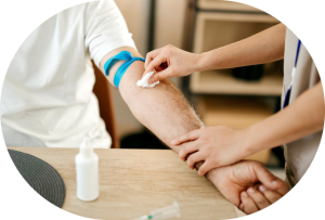
Point of care testing is a group of investigations that you would term relatively simple. These are tests such as blood tests, ECGs, echocardiography and monitors such as blood pressure monitors and ECG monitors. Typically they are done on the same day as your appointment and require no special preparation.

Summary
Dr Ravi Assomull describes what to expect from an invasive coronary angiogram. In this video, he explains why the procedure is routinely performed, how risks are minimised and when it may lead to immediate treatment.
Transcript
If you've been referred for an invasive angiogram scan, you shouldn't be worried. We really only refer our patients if we genuinely think there is a high likelihood of finding severe narrowings in the blood vessels that supply your heart. The way we work is that we ensure that your scan will be carried out by a highly experienced operator. And I work with a group of colleagues who undertake high volumes of these procedures with excellent outcomes. You will have the scan done typically in a hospital where there are backup facilities should there be any concerns or should you need any treatments like coronary angioplasty or stenting. So there's nothing here to be worried about. It's in some ways a routine procedure that gets done day in, day out across the country. And we're very comfortable in the way that we set our service up to look after you.

Summary
Dr Ravi Assomull describes the key blood tests used to assess cardiovascular risk. In this video, he explains the importance of cholesterol, metabolic markers and advanced biomarkers, alongside imaging, for a complete heart health assessment.
Transcript
Blood tests are a really important part of any assessment, including heart risks. And typically, our standard panel of blood tests will include your lipid levels. That includes your cholesterol, which is broken down into your good cholesterol, your bad cholesterol, and your triglycerides. We complement this with other factors that can be affected or show signs of potential underlying metabolic dysfunctions. So your liver function tests, your kidney function tests, your thyroid function. More recently, we started to incorporate advanced biomarkers, things like lipoprotein A, apolipoprotein A, and apolipoprotein B. However, it's important to say that blood tests aren't the be-all and end-all. You can have a normal set of blood tests or a very abnormal set of blood tests. But we have to put that in context with your clinical history, your family history, and supplement that with cardiac scans. And specifically, things like a CT coronary angiogram, which actually shows whether you've got any underlying plaque.

Summary
Dr Ravi Assomull explains how a DEXA scan provides insight into body composition and bone health. In this video, he outlines how the scan helps guide personalised exercise, nutrition and long-term health planning.
Transcript
A DEXA scan is a low radiation body composition scan that tells us about your body composition, as in how much fat and muscle you have, and also the distribution of the fat, whether it's subcutaneous or more concerningly based around organs, something we refer to as visceral fat. Additionally, DEXA scans have traditionally been used to look at your bone mineral density, how strong your bones are, and this is something particularly important in postmenopausal women, where we worry about osteoporosis. The scan itself is really very simple. It involves you lying on a bed for 10 minutes while a camera, in essence, passes over your body over two sweeps or so. There are no needles involved and the results are available pretty much immediately. The results allow us to look at your body composition and then formulate a plan for your exercise, nutrition and bone health.

Summary
Dr Ravi Assomull explains what to expect during your first cardiology consultation. In this video, he outlines how a detailed discussion and tailored investigations help identify risks and guide personalised treatment plans.
Transcript
The first cardiology assessment is really our opportunity to have an open discussion about your concerns, your family history, your lifestyle, the pressures you face, the challenges that you'd like to overcome. And then from that, we tailor a bespoke series of tests that will inform us as to what the best way of treating you is. And we also focus on your lifestyle, having an in-depth discussion on what your day involves, what your week involves, how you can potentially reconfigure things. And part of that is really getting the idea that you need to instill new habits that focus on prioritising sleep, eating well, exercising regularly. And that's something that I've done for my own family, for myself, and it's something I really enjoy working with when I see patients.

Summary
Dr Ravi Assomull describes how a stress echocardiogram works. In this video, he explains how exercise or medication is used to assess heart function under stress and identify potential issues with blood supply.
Transcript
During a stress echocardiogram our aim is to get your heart rate up to 85% of your maximum predicted heart rate and we do that in one of two ways we either get you to exercise on a treadmill or using a bike or we actually give you a medication to speed your heart up something called Dobutamine is the medication we do for that. So that's how we achieve the stress that can be physiological with exercise or pharmacological with medications such as Dobutamine. Now the key thing in a stress echocardiogram is we want to assess your heart muscle when you are at your near peak exercise or peak heart rate levels. We do this by taking pictures of the heart and what we're looking for is to see if any part of the heart starts pumping less forcefully because that may suggest that that part of the heart muscle isn't getting adequate blood supply.

Summary
Dr Ravi Assomull, a Private Cardiologist in London, explains what normal heart test results mean. In this video, he highlights how reassurance is combined with an integrative cardiology approach focused on long-term prevention.
Transcript
If the tests that we've organised for you are normal, then I think we can take a great deal of encouragement that actually you are in a good space in terms of the risk to you from your symptoms. Normally the tests we organise will reassure both you and me that the symptoms pose no risk to you. And then it's a case of trying to get on top of the symptoms with either reassurance or medications. But more importantly, we need to think about your overall health for not just the next few years, but the next few decades. And whilst this single snapshot of normal tests can reassure you, my approach is to counsel you on how to keep your heart healthy for the next few decades. And that really taps into my role as an integrative cardiologist where we focus on lifestyle, specifically sleep, exercise and nutrition.

Summary
Dr Ravi Assomull explains how a tilt table test helps diagnose causes of dizziness or fainting. In this video, he outlines how changes in heart rate and blood pressure are monitored to identify conditions such as syncope or POTS.
Transcript
A tilt table test is a useful test when we're trying to understand why people are getting dizziness or perhaps even losing consciousness. The most common cause of these symptoms are things like vasovagal syncope, fainting, POTS, which is a form of heart rate increase called postural orthostatic tachycardia syndrome. And very simply, what happens on this day when you have your tilt test is that you come in, you lie flat on a motorized table, you have an ECG attached to you as well as a blood pressure cuff. And after resting on a flat table for a matter of 10 to 15 minutes, having your heart rate, your blood pressure and your ECG recorded, you are then tilted to about 70 degrees or almost upright and kept there for about 20 minutes. And what we're interested to see is what happens to your heart rate, what happens to your blood pressure and whether we reproduce the symptoms of dizziness or perhaps even loss of consciousness. It's important to say at this stage, this is an entirely safe test. There's no risk and it can be informative in those people where we're unsure as to the cause of loss of consciousness. In some of our patients, we may actually give you a spray of a medication called GTN to see if that reproduces your symptoms.

Summary
Dr Ravi Assomull describes advanced cardiac imaging techniques such as MRI and CT scans. In this video, he explains when these tests are used to detect plaque, inflammation or structural heart disease beyond standard imaging.
Transcript
So when we talk about advanced cardiac imaging, we're typically referring to scans such as MRI, CT, or perhaps even nuclear imaging, something we use far less often nowadays due to the high radiation burden. So I'd say cardiac, MRI and cardiac CT are what I would suggest advanced imaging is represented by. And that's separate to our standard ultrasound imaging, our echocardiography, which we offer to most of our patients. Now we use advanced imaging when we need to look at specific issues. Is there any evidence of blockages in the coronary artery? So a CT coronary angiogram is considered the gold standard to understand whether there is buildup of plaque and potential blockages in the coronary arteries. And an MRI of your heart gives us a real understanding of whether there is any inflammation or scarring in the heart muscle, something called tissue characterisation, which an echocardiogram is unable to do.

Summary
Dr Ravi Assomull explains why VO2 max is an important measure of cardiovascular fitness. In this video, he outlines how improving VO2 max through exercise can significantly reduce long-term health risks and support heart health.
Transcript
Well, VO2max is an excellent measure of your cardiorespiratory fitness. So it's a way of assessing how your heart, lungs and muscle can use oxygen during exercise. And what we've learned is that for every small increase in your VO2max, something we refer to as 1MET, there's a 10 to 15% decrease in your mortality. And we know that actually if you look at the people in the elite level versus those with the lowest levels of VO2max, there's a five-fold increase in mortality in those with the lowest levels of VO2max. The really good news is your VO2max is completely trainable. You're not stuck with your current VO2max. There are exercise training protocols, utilising high-intensity interval training, where you can actually boost your VO2max and thereby increase your health span and lifespan.

Summary
The most appropriate heart tests depend on your symptoms and risk factors. In this video, Dr Ravi Assomull explains why simple tests often provide the most valuable information.
Transcript
The best screening tests to have really depend on your personal circumstances, what you're hoping to understand about your heart health. But in general, we try and keep things really simple. We start by simply taking a history, examining you and arranging blood tests and an ECG tracing as a typical baseline. Often a combination of these relatively simple assessments gives us a clear understanding of your heart health and your future risk of cardiovascular disease.

Summary
Additional tests may be required to reach a complete diagnosis. In this video, Dr Ravi Assomull explains how existing NHS results are used and when further testing is helpful.
Transcript
So when you come and see us, it may actually be the case that you don't need more tests. If you've had tests with your GP or in the NHS, we make utmost efforts to secure those results around the time of your consultation. So we can use that information without having to repeat any tests, which is obviously not something that adds any value at all. However, we also need to acknowledge that to make a full diagnosis, you may need further tests that haven't yet been provided from your previous consultations or your experience in the NHS.

Summary
Normal test results are reassuring and indicate no serious heart disease. In this video, Dr Ravi Assomull explains how symptoms can still be managed safely even when tests are normal.
Transcript
So it can be quite frustrating if you have symptoms and you have a whole load of tests and they're then told everything's normal. The way to look at it is actually good news because if your tests are normal that means there's no risk to you and that really is what matters. The focus can then switch to symptoms which can often be controlled using lifestyle modifications or medications for hopefully just a short period...

Summary
After test results are available, a follow up plan is created to address symptoms and monitoring needs. In this video, Dr Ravi Assomull explains what patients can expect after their consultation.
Transcript
Typically after we have all your results available to us, and that may be the results of tests we've organised or the results from tests you've had previously, we arrange a follow-up appointment to go through everything and formulate a plan on how to treat any symptoms you may have, and also to plan ahead of what monitoring you may need for any condition we diagnose.

Summary
Blood pressure can be measured accurately at home or in a clinical setting using an automated cuff. In this video, Dr Ravi Assomull explains correct measurement and what readings are considered normal.
Transcript
Blood pressure can easily be measured in the comfort of your own home by buying a blood pressure machine. This involves putting a cuff on your arm, pressing a button and allowing the machine to measure your blood pressure. Obviously these checks can be carried out in your GP surgery or with us in our clinic, but we tend to advise that your blood pressure is best checked at home, where reading should on average be less than 135/85.

Summary
Private cardiology tests mirror NHS investigations but are delivered differently. In this video, Dr Ravi Assomull explains how speed, convenience, and test clustering benefit patients.
Transcript
The tests we provide are identical to the tests that are available in the NHS. We don't provide anything further above and beyond what the NHS provides. However, there are some differences in the way we deliver these tests. The first thing to say is we do this quickly. We don't want you to wait and worry about the possible results of these investigations. And the second thing to say is we don't want you going backwards and forwards to have tests. We tend to try and cluster all your appointments together. So all your tests are done typically in one trip to us. Obviously, if you have a large number of tests, that may not be possible. But then we try and restrict it to times that work for you and to minimise the number of visits you make to our clinic.

Summary
Blood pressure readings include systolic and diastolic values that reflect heart function. In this video, Dr Ravi Assomull explains what these numbers mean for your cardiovascular risk.
Transcript
The two numbers in your blood pressure reading correspond to the pressure in the vascular system of your body when the heart is contracting. This is called your systolic blood pressure and is the higher of the two numbers. As well as the diastolic blood pressure, which is the pressure in your vascular system when your heart is relaxed.

Summary
An echocardiogram is a simple, non-invasive test that uses ultrasound to capture moving images of your heart. In this video, Dr Ravi Assomull explains exactly what happens during the scan, what it feels like, and how it helps assess the health of your heart valves and muscle. Learn what to expect before, during and after your appointment and how this test plays a vital role in diagnosing heart conditions.
Transcript
An echocardiogram is a safe, non-invasive and painless test that uses ultrasound to create moving pictures of the heart. This provides invaluable information about the structure and function of the heart muscle as well as of the four valves that reside within the heart. When you arrive, you'll be asked to lie on a bed, usually on your left side. A cardiac physiologist will place some gel on your chest to help transmit sound waves. Then, they will move a small handheld device called a transducer over different areas of your chest. You may feel slight pressure as a physiologist pressures the transducer against your skin, but it shouldn't be painful. During the scan, you'll hear the sound of your heartbeat and maybe ask to hold your breath briefly to get clearer images. The test usually takes 30 to 45 minutes. Afterward, you can wipe off the gel and go about your day as normal. The doctor will review the images from your echocardiogram and discuss the results with you at your follow-up appointment.

Summary
Cardiac MRI is a powerful, radiation-free imaging technique that offers detailed insights into your heart’s structure and function. In this video, Dr Ravi Assomull explains how cardiac MRI is used to diagnose conditions like cardiomyopathies, myocarditis, and coronary artery disease and walks you through exactly what to expect on the day of your scan. Learn how stress MRI, contrast agents, and high-resolution imaging help us detect hidden causes of chest pain, breathlessness, and more.
Transcript
When patients present with symptoms such as chest pain, breathlessness, palpitations or unexplained heart failure, conventional tests such as an ECG and an echocardiogram may not provide sufficient data for us to be able to fully explain what's going on. That's where cardiac MRI steps in. Cardiac MRI is considered the gold standard imaging modality to assess heart structure and function and critically to provide information on tissue characterisation. By that I mean what the heart muscle is made of. Is it scarred? Is it damaged? Is it inflamed? Cardiac MRI plays an increasing role in a majority of our patients presenting to our clinic. It is critical in helping us to understand and diagnose a range of conditions including cardiomyopathies which are a form of heart muscle disease as well as myocarditis and pericardial disease. Critically it does all of this without using any ionizing radiation whatsoever. For example, in cases of conditions such as myocarditis, cardiac MRI can detect areas of oedema and inflammation. This is something no other test can do. For that reason, cardiac MRI is considered critical in diagnosing myocarditis. Additionally, another large application is the use of stress cardiac MRI to detect possible underlying obstructive coronary artery disease. Stress cardiac MRI can detect areas where the heart isn't getting adequate blood supply. This is a condition we call myocardial ischaemia. I'd now like to tell you what actually happens on the day of your cardiac MRI. When you arrive at our imaging unit, you'll first check in and be greeted by the MRI team. They'll explain the procedure to you and ask them safety questions such as whether you've had any metal implants or devices like a pacemaker. If you are having a stress cardiac MRI, they will ensure that you have not had any caffeine for 24 hours. Next, you'll change the hospital gown and remove any metal objects like jewelry or belts. A small cannula will usually be placed in a vein in your arm as most cardiac MRIs involve injecting a contrast dye called gadolinium. This helps highlight your heart structures. You'll then lie on a flat table that slides into the MRI scanner which looks like a large tunnel. It's important to stay as still as possible during the scan to get clear images. The scan is painless but the machine can be noisy. You'll hear loud tapping or humming sounds and you'll be given headphones or earplugs. You'll be asked to follow simple breathing instructions like hold your breath for a few seconds at a time. The entire process can take around 30 to 60 minutes. If you're having a stress MRI, a doctor will inject a medication called adenosine that speeds your heart up for four minutes. You will feel like you're exercising for those four minutes before the effects of the adenosine subside. Once it's done, you'll be monitored briefly after the contrast injection and then you can go home the same day and resume normal activities. The results are reviewed by a cardiologist and we will discuss the findings with you at a follow-up appointment. I hope you can now see that cardiac MRI plays a critically important role in managing patients with cardiovascular disease.

Summary
In this video, we walk you through what it’s like to wear a 24-hour blood pressure monitor: a simple, painless and highly effective way to get a full picture of how your blood pressure behaves throughout the day and night. Dr Ravi Assomull’s team shows you how the device is fitted and what to expect during the monitoring period. This test helps identify hidden hypertension and improves long-term heart health.
Transcript
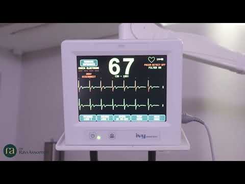
In this video we demonstrate the simple process of getting fitted with a 24-hour blood pressure monitor. A member of our team will explain everything and show you the monitor. It's smaller and lighter than you might expect. First, a comfortable cuff is placed around your upper arm. This is typically your non-dominant arm. It works just like the one you use during a blood pressure check. The cuff connects to a small machine that you wear around your waist. It's easy to carry and doesn't get in the way. The monitor will automatically check your blood pressure at regular times throughout the day and night. You don't have to do anything. Typically the cuff inflates every half hour during the day and every hour during the night. You'll go about your usual activities but it's best to avoid vigorous exercise and keep the monitor dry. After 24 hours you'll return the monitor so we can review your results and get a complete picture of your blood pressure. It's a simple and important step towards protecting your heart health. Know your numbers. Protect your heart.

Summary
Considering a CT coronary angiogram or been referred for one? In this video, consultant cardiologist Dr Ravi Assomull explains what the test involves, who it’s for, and how it helps assess your risk of heart disease. Learn how the scan detects calcium build-up, artery narrowing, and dangerous soft plaque that could lead to heart attacks. Dr Assomull also explains how modern scanners minimise radiation exposure.
Transcript
If you have been referred for or considering having a CT coronary angiogram, this video provides you with a detailed overview of the process, the benefits and the risks involved. A CT coronary angiogram is a detailed imaging modality that focuses on the blood vessel supplying your heart. We refer to these as the coronary arteries. The coronary arteries supply the heart muscle with oxygen-rich blood. A CT coronary angiogram is particularly valuable in individuals who have been experiencing symptoms such as chest pain or breathlessness and in these particular individuals it is critical to rule out blockages in the heart arteries. However, more recently these scans are being used in individuals without any symptoms but in whom we suspect that there is an increased risk of cardiovascular disease. This includes individuals who have a family history of heart disease, who have diabetes, who smoke, who have other cardiovascular risk factors such as high blood pressure and high cholesterol. There are two distinct phases in having a CT coronary angiogram. The first phase involves no contrast and detects calcified plaques. These are fatty deposits that have become hardened over time. These calcium deposits serve as a marker of atherosclerosis and provide an individualised calcium score. Your calcium score is highly predictive of your future risk of cardiovascular disease. We can use your score and compare it to individuals of the same age and gender and rank you using a percentile ranking. For example, a 50th percentile score indicates an average calcium level for your demographic, while a 99th percentile score suggests a significantly elevated risk of a future cardiovascular event. What a calcium score doesn't tell us is if there are any narrowings of the coronary arteries. To do this, we inject contrast which is the second part of the scan. When we inject contrast, we can see if there are any narrowings of the coronary arteries and critically if there is any soft fatty plaque. Now it's important to know about this as it is the soft fatty plaques which are more prone to rupture and cause heart attacks. Now let's talk about radiation. This is clearly a concern. However, we can reassure you that we use the latest generation scanners which have a fraction of the radiation exposure compared to the initial scanners we used to use. For example, our scanners will acquire all the images needed in a single heartbeat. So how much radiation do we expose you to? Well, let's put this in perspective. The radiation dose emitted by our latest generation CT coronary angiogram scanner is equivalent to the dose you would absorb if you lived in Cornwall for six months.
Summary
In this video, Dr Ravi Assomull walks you through exactly what to expect on the day of your CT coronary angiogram, from preparation and scanning to aftercare. Understand the step-by-step process, how we ensure your safety, and why this non-invasive scan is such a powerful tool for assessing your heart health. Learn how to prepare for the scan and what it tells us about your coronary arteries.
Transcript
The video you're about to watch tells you everything you need to know on the day of your CT coronary angiogram. So there are several things that we need you to do before the scan. The key here is that you have to avoid caffeine on the day of the actual study. It's also important that you refrain from eating for six hours prior to the scan. Once you come into our unit we'll ask you to change into a hospital gown and remove any metal objects such as jewellery. During the actual scan the first thing we do is put you on our motorised bed and get you in the right position. This allows the bed to move into the CT scanner which resembles a large donut. Prior to doing that we put some electrodes onto your chest so that we can monitor your heart rhythm. This allows the scanner to acquire the images at an appropriate time on your heart cycle. Once that's done and you've had the first stage of the scan which does not involve any contrast, we move on to the second stage where a contrast dye is administered via the intravenous line that we've put in place. Once that's done you experience a warm sensation or perhaps even a metallic taste in your mouth. The scanner takes the images and it is critical that you keep as still as possible. The other thing that we ask you to do is hold your breath for a few seconds at a time during the study. The actual scan itself takes just 5 to 15 minutes but the overall procedure once we factor in paperwork, getting you dressed and getting you changed again may take up to 30 minutes. In a proportion of patients, particularly those who are understandably anxious, your heart rate may be higher than usual. It's important to get your heart rate low for the scan so we give those patients intravenous beta blockers which are medications that reduce your heart rate. After the scan is complete, you can resume normal activities immediately. It's important to drink plenty of water to help flush out the contrast die. Once the scan is complete, the images are passed on to cardiologists and radiologists who work together to provide a detailed report on the findings of your scan. I hope you now realise that a CT Colony Angiogram is a safe and highly informative diagnostic tool that provides crucial insights into your heart health without the need for invasive procedures.
Summary
If you’ve been referred for an ambulatory ECG monitor, this video explains everything you need to know. Dr Ravi Assomull walks you through the entire process — from preparation and fitting to wearing the monitor and returning it.
Transcript
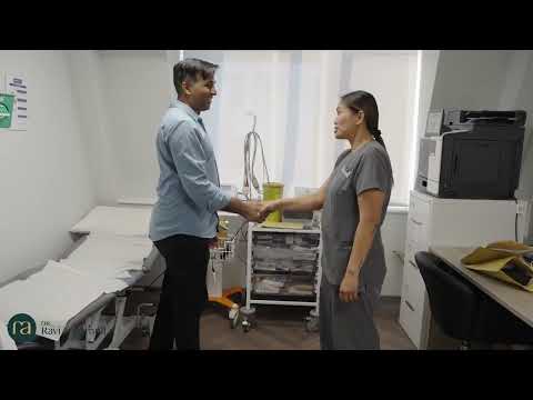
If you're watching this video, it's likely you've been referred to wearing an ambulatory ECG monitor. In this video, we walk you through what to expect on the day of your appointment. An ECG monitor or patch is a small wearable device that continuously records your heart's electrical activity, usually over 24 hours to 14 days. It helps your doctor see how your heart rhythm behaves as you go about your normal routine. On the day of your appointment, don't use body moisturisers, perfumes or powders as they can stop the electrodes from sticking properly. We may need to shave a small area of chest hair to help the electrodes attach securely. You can do this yourself the night before or we'll do it for you during the appointment. If you'd like a chaperone, just let us know in advance. When you arrive, you'll be asked to remove your clothing from the waist up. A cardiac physiologist will explain the process and prepare your skin using an alcohol wipe and a special pad. The electrodes will then be applied to your chest and the Holter monitor will be connected. If you're wearing it for more than 24 hours, we'll show you how to change the electrodes yourself. The whole process takes around 15 minutes. While wearing the monitor, you can carry on with your daily activities including showering. Just keep showers short and avoid getting soap or direct spray on the monitor. No baths or swimming though. Make sure to keep a diary of your symptoms and activities. This helps your doctor match any events to what your heart was doing at that time. If the patch starts to peel, press it back down, especially morning and night. If it won't stick, you can use medical tape to secure it. When your monitoring period is over, you can remove the patch yourself, then just drop off the monitor and your diary in the clinic. Your results will be analysed in a day or two, ready for us to review at your follow-up appointment.
Explore and learn about the various heart tests and diagnostic options we provide.
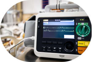
Electrocardiogram (ECG)
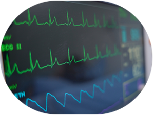
An ECG can tell us your heart’s rate, rhythm, and electrical activity, and it can pick up any irregular beats.
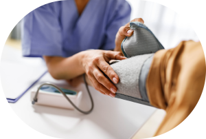
Health MOT
This overall check, including blood pressure checks, can detect any signs of a health issue.
24-hour blood pressure monitor
Observing your blood pressure levels over 24 hours can show us how your heart is performing daily.
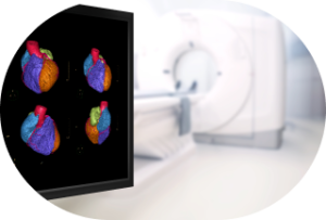
Advanced imaging
We offer advanced imaging scans to provide further insight into how your heart works and highlight issues with valves and vessels.

Ambulatory ECG monitor
Ambulatory ECG monitors can help record your heart’s activity during daily tasks.
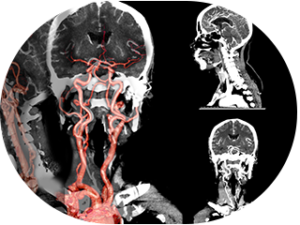
Angiogram scan
A cardiac angiogram can visualise your heart’s blood vessels.We can use precise imaging to help detect blockages and assess your cardiovascular health.
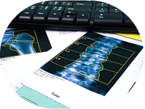
Bone density (DEXA) scan
Our specialised DEXA scanning evaluates bone density risks linked to cardiovascular concerns.
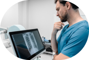
Chest X-ray
Get insights into heart chest x-ray tests. Learn about the procedure, its importance in detecting heart conditions, and what patients can expect.
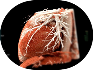
CT coronary angiogram scan
A CT coronary angiogram can help us examine heart arteries and assess your cardiovascular health precisely.
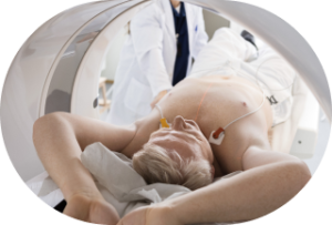
Echocardiogram
Discover what an echocardiogram is, how it works, and why we use this ultrasound test to assess heart health and diagnose cardiac conditions.
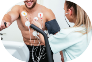
Exercise ECG
Sometimes, your heart’s activity while you’re exercising can give us insights into your condition.
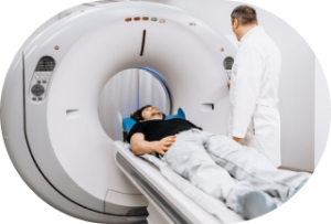
Heart MRI scan
Comprehensive heart imaging through advanced MRI technology. We provide detailed scans to evaluate your cardiac health and detect potential heart conditions.
Initial assessment
To determine whether or not your concerns are a sign of a heart issue, we will perform some initial tests.
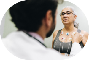
Stress echocardiogram
Concerned about your heart performance? Our stress echo test reveals how your heart functions during exercise.

Tilt table test
A tilt table test can help us determine why you might be fainting. Read more to find out how the test is performed and how to prepare beforehand.
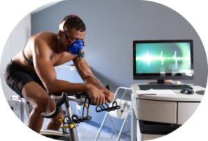
VO2 max testing
Trust us for comprehensive VO2 max testing. We can evaluate your heart’s efficiency and exercise capacity to provide personalised treatment.
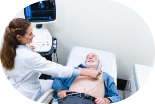
Point of care diagnostics
It’s always important to thoroughly investigate signs of a heart concern. These diagnostic tests are key to discovering what may be affecting your heart.

Transoesophageal echo (TOE)
Close-up ultrasound of the valves and chambers from the food pipe - exceptional detail when standard scanning needs a closer look.

Implantable loop recorder (ILR)
A tiny implanted monitor that watches every heartbeat for up to four years - catching the rhythm problems short-term monitors miss.
Explore the specialist heart treatments and procedures we provide.
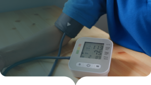
Cardioversion
A brief, planned procedure that resets atrial fibrillation or flutter back to normal rhythm with a carefully controlled electrical impulse.

Catheter ablation
Keyhole treatment for AF, flutter, SVT and VT that targets the abnormal rhythm at its source - often curing it altogether.

Coronary angioplasty & stenting
A keyhole procedure to open narrowed coronary arteries, relieve angina and restore healthy blood flow to the heart muscle.

Implantable defibrillator (ICD)
A small implanted device that continuously guards against life-threatening heart rhythms - treating them within seconds.

Pacemaker implantation
The definitive treatment for slow heart rhythms, blackouts and heart block - and the basis of resynchronisation therapy for heart failure.

TAVI (keyhole valve replacement)
Keyhole replacement of a severely narrowed aortic valve through a small puncture at the groin - without open-heart surgery.
Book your consultation with Dr Ravi Assomull today
Looking after your heart is the most important thing you can do to improve your longevity and quality of life in the long term.
We’re here for you during your journey to better heart health. We provide tests and management strategies to help identify what might be wrong and where you can improve your lifestyle to reach prime heart health.

Book an appointment today to speak to our expert Integrative Cardiologist, Dr Ravi Assomull, about your heart concerns.
You can email us at: enquiries@cardiologist.london
Or call us at: 020 3576 2885